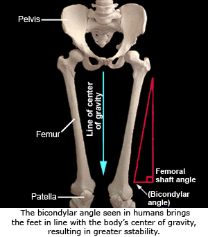
 A critical adaptation for efficient bipedalsim relates to the need to keep the body’s center of gravity balanced over the stance leg during the stride cycle. Birds solved this issue by having the entire leg (from the hips all the way to the feet) as close as possible to the body’s center line. In humans, whose hips are wide apart, the shaft of the femur is angled so that the knee is closer to the body’s midline than the hips. This angle is called the bicondylar angle, and the resulting knee joint is referred to as a valgus knee. The effect is to bring the knees closer together, placing the feet directly below the center of gravity.
Compared to modern humans, an ape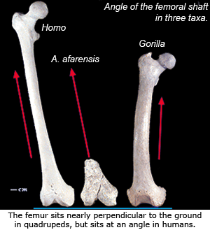 femur is almost vertical within a horizontal plane. In quadrupeds the positioning of the center of gravity during locomotion is less critical since the quadruped is usually supported by 2 or more legs during the stride cycle rather than just 1 as with humans. Australopiths have a human-like bicondylar angle9,18.
A result of the femur’s bicondylar angle pulling the knees inward is that the tibia stands almost parallel with the body’s center of gravity. Unlike the femur, the tibial shaft lies at a right angle to its proximal surface. Also of note, the human medial and lateral proximal articular condyles (i.e., the flattened surfaces on the top of the tibia that articulate with the femur) are relatively larger and elongated anteroposteriorly (i.e., longer front to back) compared to quadrupeds. The comparatively larger lateral proximal condyle (also seen on the femur and helps to create the bicondylar angle) is an adaptation to increased weight transfer through the femur and into the foot. In addition, modern human condyles are more concave and elliptical in shape to accommodate the elliptical femoral distal condyles. Quadrupeds tibial condyles appear relatively spherical and are more convex. The elliptical shape in humans helps to lock the knee in place and create straight-forward forward leg movement9.
A chimpanzee's tibia retains smaller lateral proximal condyles, and may exhibit an obtuse angle between the tibial proximal surface and the shaft. The autralopith tibia has a nearly right angle between the shaft and the proximal surface.