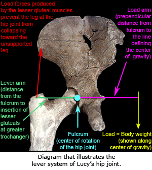
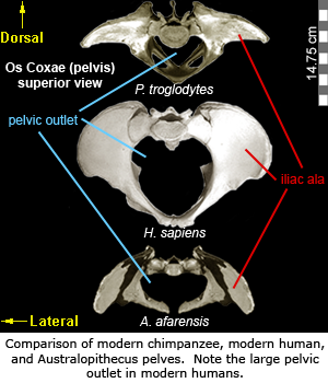
 The gluteus medius and gluteus minimus muscles originate on the dorsal side of the ilium and insert on the greater trochanter of the femur. Their actions are critical to propulsion and stability while walking. Since bipedalism requires special adaptations, the orientation (and thus the function) of the gluteal muscles, is different in bipedal humans and quadrupedal apes. In apes, the flat portion of the iliac ala is roughly parallel with the plane of the back, while in humans the iliac ala is shifted laterally and flares more on the sides. This relatively lateral orientation of the alae in humans abducts (i.e., move away from the body) the hip joint. In turn, the gluteal muscles act to stabilize the area by preventing the hip on the supported side (the standing leg) from collapsing toward the unsupported side (the swinging leg). In apes, these muscles are attached relatively dorsal (i.e., more toward the back and less on the sides) and act as hip extensors, which move the leg backward when the primate takes a step9.
 The australopith pelvis exhibits widely flaring iliac ala. This flare is a critical component of the lever system of the hip and acts to increase the mechanical advantage of the lesser gluteals by increasing their lever arm. However, the lateral flare of the australopith ala is more pronounced than typically seen in modern humans. Shape similarities between the australopith and modern human pelves indicate that Australopithecus was fully bipedal. However, the unique morphology seen in australopiths suggests the species did not utilize the modern gait seen in later Homo9.
The modern human pelvis has relatively larger hip joints and larger pelvic outlet relative to australopiths or modern apes. These differences appear to be a compromise between two functional needs: 1) efficient bipedalism; and 2) allowing enough space for wide shouldered, large brained infants to pass through the birth canal.
 In bipeds, the hips support and balance the weight of the torso during locomotion. However, as the size of the pelvic outlet increased, the hip joints were repositioned relatively further from the center line of the body. As a result, more force is exerted on the hip joint as the joint (acetabulum and femoral head) moves further away from the body’s center of gravity, and thus affects stability as the weight of the torso presses downward toward the middle of the body. This issue is resolved through several adaptations in the pelvis and femur. In the pelvis, an enlarged hip joint allows more stress to be absorbed and accommodates a larger femoral head9,16.
eFossils is a collaborative website in which users can explore important fossil localities and browse the fossil digital library. If you have any problems using this site or have any other questions, please feel free to contact us.
Funding for eFossils was provided by the Longhorn Innovation Fund for Technology (LIFT) Award from the Research & Educational Technology Committee (R&E) of the IT governance structure at The University of Texas at Austin.
